 Skip to Content

How Eustachian Tube Balloon Dilation is Performed
If you suffer from Chronic Eustachian Tube Dysfunction, you may find relief from Eustachian Tube Balloon Dilation.
In the past, individuals with ETD were treated primarily with repeated ear tubes. However, a new minimally invasive surgical technique uses a balloon to dilate this important passageway and remodel the cartilage, allowing patients to have longer, more effective relief.
Before the procedure
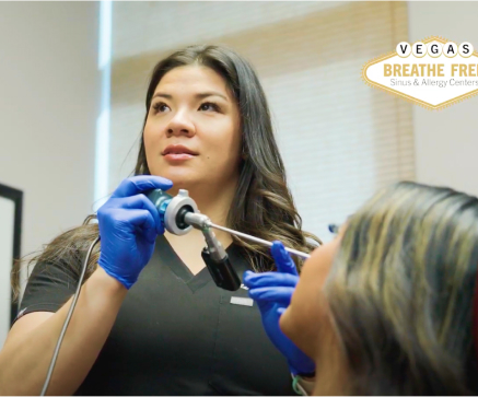
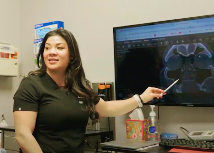
Your surgeon will place a small endoscope — a thin tube with a camera and light — through the nasal opening to examine the nasal cavity and look for any potential causes of ETD or assess for anatomical issues that may impact the treatment setting for you. Examples of these include ruling out sinus infections, extent of acid reflux, severity of allergic rhinitis, presence of septal deviations, etc.

On the day of your procedure
An endoscope is introduced into the nose with the deflated Eustachian tube balloon. The device is guided through the nasal passageway until it reaches the eustachian tube opening in the back of the nose. Under direct visualization, the balloon is then inserted into the Eustachian tube and inflated to dilate the blocked tube — similar to the way a stent is used to open up a clogged artery. The balloon is kept inflated for about two minutes helping to remodel the Eustachian tube cartilage and flatten out any inflammation. The balloon is then deflated and removed from the Eustachian tube.In the case that both Eustachian tubes are affected, the procedure can be performed on the opposite side as well.
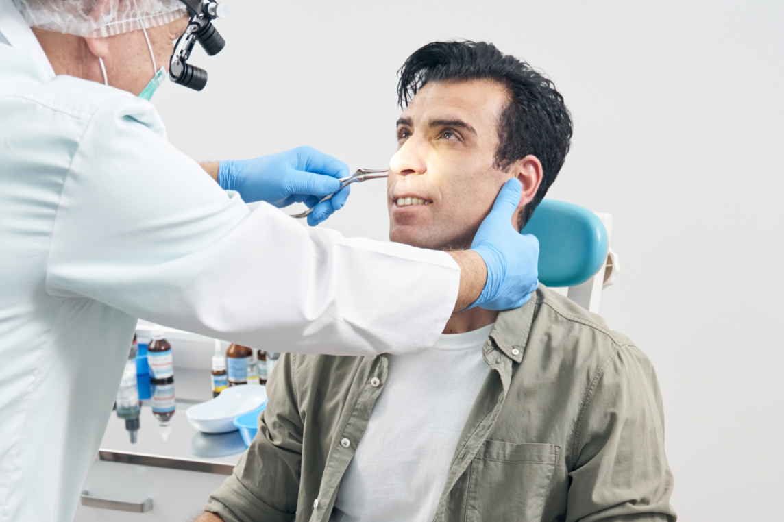
Eustachian Tube Balloon Dilation can be performed safely and effectively under either local anesthesia in the office setting or general anesthesia in the operating room. When done in the clinic, the majority of the procedure is numbing the nose with a variety of sprays and topical medications. Ultimately, once the nose is anesthetized, the procedure is well tolerated with minimal discomfort.
To perform the procedure
The physician must be able to move the balloon freely through the opening of the nostril and into the eustachian tube. Individuals with anatomical issues, such as a severely deviated septum or nasal polyps, may benefit from other surgical approaches in the operating room. After the initial evaluation, your surgeon will help guide which treatment setting would be best considering patient preference, anatomical abnormalities and other factors.
Benefits of Eustachian Tube Balloon Dilation
For decades, the gold standard of treatment for patients with chronic eustachian tube dysfunction has been repeated placement of ear tubes. However, these tiny plastic cylinders that are surgically placed in the ear can fall out over time and long-term use can lead to perforation and often necessitates dry ear precautions. Since Eustachian Tube Balloon Dilation was introduced in 2010, numerous studies have shown the procedure has several distinct benefits.
Candidates for Eustachian Tube Balloon Dilation
Individuals who would benefit from the procedure suffer from chronic ETD or otitis media, otherwise known as recurrent ear infections. These patients typically have had several prior ear tube surgeries, report a constant feeling of pressure or fullness in the ear.
The medical management of underlying conditions is critical prior to surgery. Before anyone considers balloon dilation, allergies, sinonasal disease and laryngopharyngeal reflux must be well controlled. Many of these conditions if not treated may lead to less-than-optimal outcomes and continued symptoms.


Follow-up Care and Recovery Time for Eustachian Tube Balloon Dilation
Eustachian Tube Balloon Dilation is a simple, safe, and effective procedure. Patients typically return to work within a day or two. Some individuals may experience mild soreness or congestion. Patients should expect no visible bruising or swelling. Post-operative nasal sprays are generally prescribed both for comfort and to help moisturize the nasal passageways.
As with any surgical procedure there may be risks associated with Eustachian Tube Dilation. Some of the most common risks include nose bleeds, infection, trauma to the Eustachian tube, over opening of Eustachian tube (patulous Eustachian tube) and lack of symptom improvement.
After one week, your surgeon may recommend an exercise known as the Valsalva maneuver. They will teach you to hold your nose, close your mouth, and blow out — just as you would clear your ears on a plane. The Valsalva maneuver forces air through the Eustachian tube and can help relieve pressure buildup and with remodeling the newly treated cartilage.